
The Department of Medicine and Surgery possesses departmental instruments capable of performing high-tech applications. These instruments are currently operational and freely accessible to all researchers within the Department.
Plate reader INFINITE F NANO
 The instrument is located in Laboratory B-P04-U39 (Building B, 4th floor, room 39).
Reference Researchers:
Maria Laura BELLADONNA This email address is being protected from spambots. You need JavaScript enabled to view it.
Chiara SUVIERI This email address is being protected from spambots. You need JavaScript enabled to view it.
Sofia ROSSINI This email address is being protected from spambots. You need JavaScript enabled to view it.
F nano+ is the model from the Infinite 200 PRO series based on filters for readings of:
A. Absorbance
B. Top fluorescence (including QuBit, Quant-IT, PicoGreen, and FRET assays)
C. Bottom fluorescence (ideal for adherent cells, with a lid to maintain sterility if needed)
D. Time-resolved fluorescence: supports both TRF and TR-FRET assays. F nano+ is certified for HTRF® readings.
The reader comes standard with temperature control and plate shaking. It also allows multiple readings per well, ideal for heterogeneous sample distribution.
The Infinite readers are compatible with standard SBS-format microplates from 6 to 384 wells and with the NanoQuant Plate™ for absorbance measurements in 2 µl for 1-16 samples per batch (filters must be ordered separately). Well volumes depend on the plate specifications: for example, with Corning CLS3540 plates (and similar), fluorescence can be read for samples with volumes between 5 and 40 µl, for 1 to 384 samples at a time. (Operating Funding 2024).
Workstation Dell PRECISION 5860
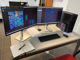 Location of the equipment: Building D, ground floor
For the use of the above-mentioned equipment, please refer to the researchers listed below:
Reference Researchers:
Giuseppe NOCENTINI This email address is being protected from spambots. You need JavaScript enabled to view it.
Luigi CARI This email address is being protected from spambots. You need JavaScript enabled to view it.
The workstation, based on a next-generation Xeon platform and equipped with 256 GB of RAM and a GPU featuring Nvidia CUDA architecture, is designed for processing large volumes of data that require high-performance computing and complex simulations through the use of specialized software.
Specifically, the workstation is intended for:
- Processing high-parameter flow cytometry data using dimensionality reduction algorithms;
- Processing confocal microscopy images (image analysis and deconvolution);
- Processing transcriptomic data (bulk RNA-seq and single-cell RNA-seq);
- Processing spatial transcriptomics and proteomics data (machine learning–based clustering). (Fondo Funzionamento 2024)
ThermoABI QuantStudio 7 Flex Real-Time PCR (with 384-well thermal block)
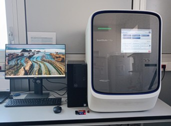 Location of the equipment: Building B, 3rd Floor
For the use of the above-mentioned equipment, please refer to the researcher listed below:
Reference Researcher:
Michele BIAGIOLI This email address is being protected from spambots. You need JavaScript enabled to view it.
The ThermoABI QuantStudio 7 Flex Real-Time PCR system is a high-performance platform for the real-time quantification of nucleic acids, suitable for both genomic and transcriptomic applications. Equipped with interchangeable thermal blocks, including a 384-well format, it enables high-throughput analysis with high precision and significantly reduced cost per sample. The instrument supports a wide range of real-time PCR assays, including TaqMan® and SYBR® Green chemistries, and is ideal for genotyping studies and gene expression analysis through mRNA transcript quantification. Integration with advanced data analysis software ensures accurate, reproducible, and robust results, even in complex experimental designs (Fondo Funzionamento 2024).
Phenocycler/Fusion for Spatial Microscopy
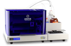
The equipment is located in Building A, on floor -2. To use the equipment, it is necessary to refer to the researcher in the list below.
Reference Researchers:
Francesca FALLARINO This email address is being protected from spambots. You need JavaScript enabled to view it.
Luigi CARI This email address is being protected from spambots. You need JavaScript enabled to view it. + 39 3481234845
Giulia MENCARELLI This email address is being protected from spambots. You need JavaScript enabled to view it. +39 3479536150
Monia BILLI This email address is being protected from spambots. You need JavaScript enabled to view it. +39 3385800189
PhenoCycler-Fusion 2.0 is a next-generation, ultrahigh-plex proteomic spatial biology system that combines automated fluidics and high-speed imaging to produce high-resolution, multi-biomarker images of tissue sections. It can image up to 1 million cells in 10 minutes, enabling comprehensive, unbiased single-cell spatial phenotyping across entire slides.
This powerful platform supports cancer research (tumor microenvironment and immune infiltration), immunology (immune responses and immunotherapies), neuroscience (neural circuits and brain immune interactions), and infectious disease studies (pathogen distribution and host responses), enhancing both pre-clinical and clinical research.
Visium CytAssist della 10x Genomics
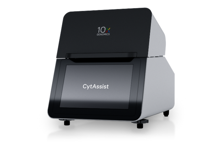
The equipment is located in Building A, on floor -2. To use the equipment, it is necessary to refer to the researcher in the list below.
Reference Researcher: Francesca FALLARINO This email address is being protected from spambots. You need JavaScript enabled to view it.
The Visium CytAssist by 10x Genomics is an innovative tool for the optimal transfer and alignment of samples on supports dedicated to spatial gene expression analysis. This technology allows the combination of transcriptomic data with histological information, improving the accuracy and reliability of studies on spatial transcriptomics, molecular biology, and personalized medicine. Integration with advanced imaging platforms, such as the Phenocycler-Fusion, expands the possibilities for multi-omics analysis in translational research and advanced diagnostics.
Confocal microscope with spinning disk
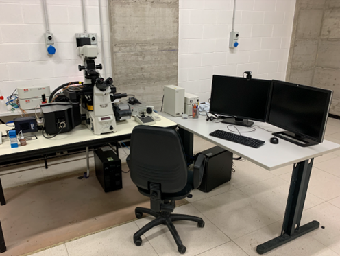
The equipment is located in the Imaging Laboratory, Building A, floor -2.
To use the aforementioned instrumentation, it is necessary to refer to the Researchers listed below, as they have been properly trained through a Training Course.
Reference researchers:
Marilena CASTELLI This email address is being protected from spambots. You need JavaScript enabled to view it.
Giada MONDANELLI This email address is being protected from spambots. You need JavaScript enabled to view it.
Marina BELLET This email address is being protected from spambots. You need JavaScript enabled to view it.
Monia BILLI This email address is being protected from spambots. You need JavaScript enabled to view it.
Martina BORDONI This email address is being protected from spambots. You need JavaScript enabled to view it.
Eleonora PETITO This email address is being protected from spambots. You need JavaScript enabled to view it.
Sara CHIAPPALUPI This email address is being protected from spambots. You need JavaScript enabled to view it.
The fluorescence microscope configured for FREET-TIRF methodology has recently been equipped with a Confocal Spinning Disc image acquisition and processing system, model X-Light V2 LFOV, manufactured by CrestOptics for high-resolution Nikon microscopes and up to 7 wavelengths (Basic Research Fund 2020). Additionally, the connection to the tabletop incubator allows for time-lapse acquisition.
Nanosight
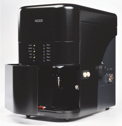
The instrument is located in the Cell Biology Laboratory, Building B, 4th Floor.
Reference researchers:
Giada CERROTTI This email address is being protected from spambots. You need JavaScript enabled to view it.
Giorgia MANNI This email address is being protected from spambots. You need JavaScript enabled to view it.
Francesca RIUZZI This email address is being protected from spambots. You need JavaScript enabled to view it.
Ilaria BELLEZZA This email address is being protected from spambots. You need JavaScript enabled to view it.
Luisa PASCUCCI luisa.pascucci@unipg,it
Maria Teresa PALLOTTA This email address is being protected from spambots. You need JavaScript enabled to view it.
Rita ROMANI This email address is being protected from spambots. You need JavaScript enabled to view it.
Sandra BURATTA This email address is being protected from spambots. You need JavaScript enabled to view it.
The NS300 NanoSight instrument utilizes the Nanoparticle Tracking Analysis (NTA) technique for the characterization of nanoparticles in solution ranging in size from 10nm to 2000nm. Both the size and concentration of nanoparticles can be measured. Additionally, the instrument allows for the identification of nanoparticles through specific immunofluorescent labeling.
Izon Automatic Fraction Collector V2
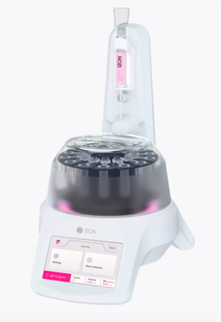
The instrument is located in the Cell Biology Laboratory, Building B, 4th floor.
Reference Researchers:
Rita ROMANI This email address is being protected from spambots. You need JavaScript enabled to view it.
Francesco FABI This email address is being protected from spambots. You need JavaScript enabled to view it.
It is an advanced device designed for the automatic fractionation of liquid samples based on specific preset parameters. It is commonly used in research laboratories and chemical and biological analysis to simplify, separate, and collect specific components of a liquid mixture precisely and efficiently based on their sizes.
The device features an intuitive user interface that allows operators to easily set desired parameters and monitor the progress of the fractionation process for accurate and reproducible fractionation of liquid samples in research and development applications (Operating Fund 2022).
Invitrogen Countess™ 3 Automated Cell Counter Starter Package
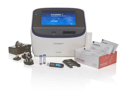
The instrument is located in the Immunology - Gastroenterology Laboratory, on the 4th floor of Building B.
Reference Researchers:
Gabrio Bassotti This email address is being protected from spambots. You need JavaScript enabled to view it.
The instrument is a "cell counter" equipped with an advanced machine learning algorithm, reinforced optics, fully automated focusing and illumination, and image analysis software for evaluating cell cultures. It counts live and dead cells (Trypan Blue), identifies cells in clusters, measures vitality, and reports the average cell size with just one touch in about 20 seconds. It can be used with disposable or reusable slides. It features a touchscreen display, WiFi connectivity, and USB connectivity (Operating Fund 2022).
Ibright CL1500 System
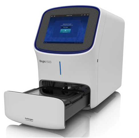
The instrument is located in the MyoLab, Building D, 1st floor.
Reference Researchers:
Francesca RIUZZI This email address is being protected from spambots. You need JavaScript enabled to view it.
Gugliemo SORCI This email address is being protected from spambots. You need JavaScript enabled to view it.
The iBright CL1500 System is an instrument for the documentation of gels and membranes in electrophoresis with high performance and safety features, equipped with powerful hardware and advanced automatic technologies (Operating Fund 2022).
Centrifuga SL1/SL1R /h5>
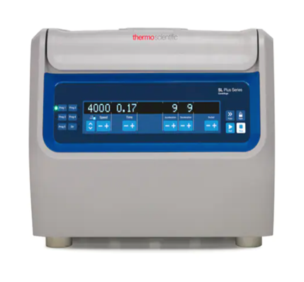
It s is located in the Cytogenetics and Molecular Biology Laboratory at C.R.E.O (Centro Ricerche Emato-Oncologiche), Santa Maria della Misericordia Hospital, Block R, Floor +2, P.le Menghini 8/0-06132, S. Sisto (PG).
Reference Researchers:
Roberta La Starza This email address is being protected from spambots. You need JavaScript enabled to view it.
This centrifuge is equipped with an easy-to-use keypad and a high-contrast LCD display. It allows for quick and safe switching between 14 unique rotor options with the tool-free Auto-Lock rotor exchange. This compact centrifuge has a capacity of up to 1.6 liters and can spin up to 76 tubes of 5/7 mL and 16 conical tubes of 50 mL with biocontainment, making it ideal for routine applications (Operating Fund 2022).
Cappa biologica bioactiva one plus 150
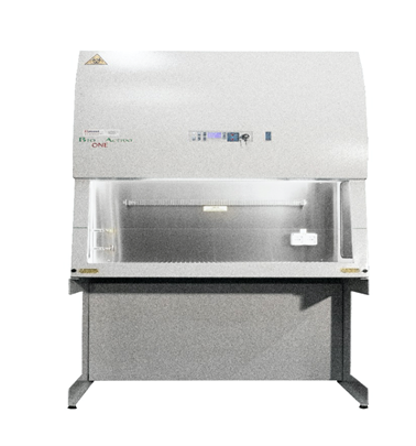
The instrument is located in the Genetics Laboratory, Educational Center, 1st floor.
Reference Researchers:
Antonio Orlacchio This email address is being protected from spambots. You need JavaScript enabled to view it.
The vertical laminar airflow sterile hood with HEPA filter ensures that 70% of the air is recirculated and 30% is expelled into the environment. Suitable for handling cells and pathogens with low to medium biological risk (Operating Fund 2022).
Co2 Incubator 150L
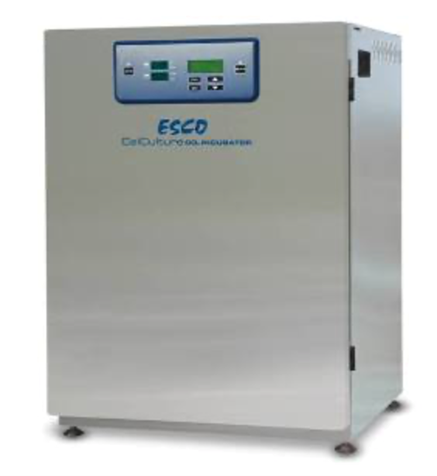
The instrument is located in the Molecular Microbiology Laboratory, Building D, 3rd floor.
Reference Researchers:
Roberta Spaccapelo This email address is being protected from spambots. You need JavaScript enabled to view it.
Alicia Yoke Wei Wong This email address is being protected from spambots. You need JavaScript enabled to view it.
The 150L CO2 incubator for cultivating and maintaining cells or tissues under controlled conditions on 3 shelves has the following features:
- External touchscreen display for monitoring and settings
- High-performance heaters surrounding the chamber for even temperature distribution
- Internal fan contributing to uniform diffusion of temperature, CO2, and humidity
- Internal glass door and stainless steel interior
- Internal tray for moisture recovery, with water level sensor
- 90°C decontamination cycle that simplifies sanitization, avoiding autoclaving and the use of toxic chemicals
- Thermal conductivity (TC) CO2 sensors, placed directly inside the chamber for precise and long-lasting control (Operating Fund 2022).
OLYMPUS BX51 Fluorescence Microscope with DP75 Camera and cellSens 4.2 software
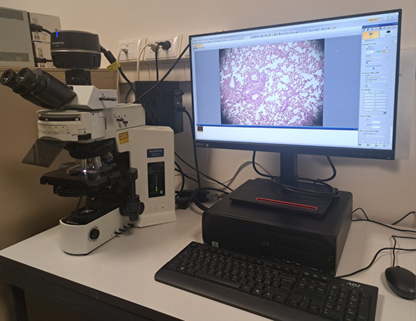
The instrument is located in the General Pathology Laboratory, Building C, 4th floor.
Reference Researchers:
Marina Maria Bellet This email address is being protected from spambots. You need JavaScript enabled to view it.
The OLYMPUS BX51 fluorescence microscope is an advanced system ideal for observing biological samples in transmitted and reflected light with fluorescence capability. The fluorescence module allows sample illumination with lights of different wavelengths to excite fluorochromes and obtain high-resolution and high-contrast fluorescent images. It is equipped with a DP75 digital camera directly connected to the microscope for capturing digital images and is designed to acquire high-quality and high-resolution images thanks to the cellSens 4.2 Acquisition software that allows image acquisition and analysis. The software offers a variety of options for real-time image acquisition, allowing the user to capture still images or time-lapse sequences and provides tools for image analysis, including quantitative measurements and annotations (Operating Fund 2022).
Optima™ L-60 Ultracentrifuge
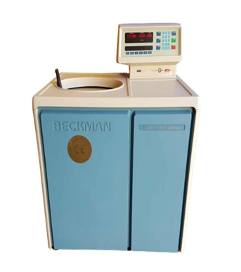
The instrument is located in the General Pathology Laboratory, Building C, 4th floor.
Ricercatori di Riferimento:
Rita ROMANI This email address is being protected from spambots. You need JavaScript enabled to view it.
The Beckman Optima™ L-60 ultracentrifuge is designed with a microprocessor-based system that ensures maximum productivity in the shortest possible time. It has a vacuum induction drive with controlled frequency that eliminates friction and increases performance. Additionally, the soft acceleration/deceleration profiles of the L-60 allow gentle handling of samples. Other features of the Beckman Optima™ L-60 ultracentrifuge include diagnostic displays that recognize condition errors, a temperature control range from 0 to 40°C, and an automatic restart system in case of power loss (Operating Fund 2022).